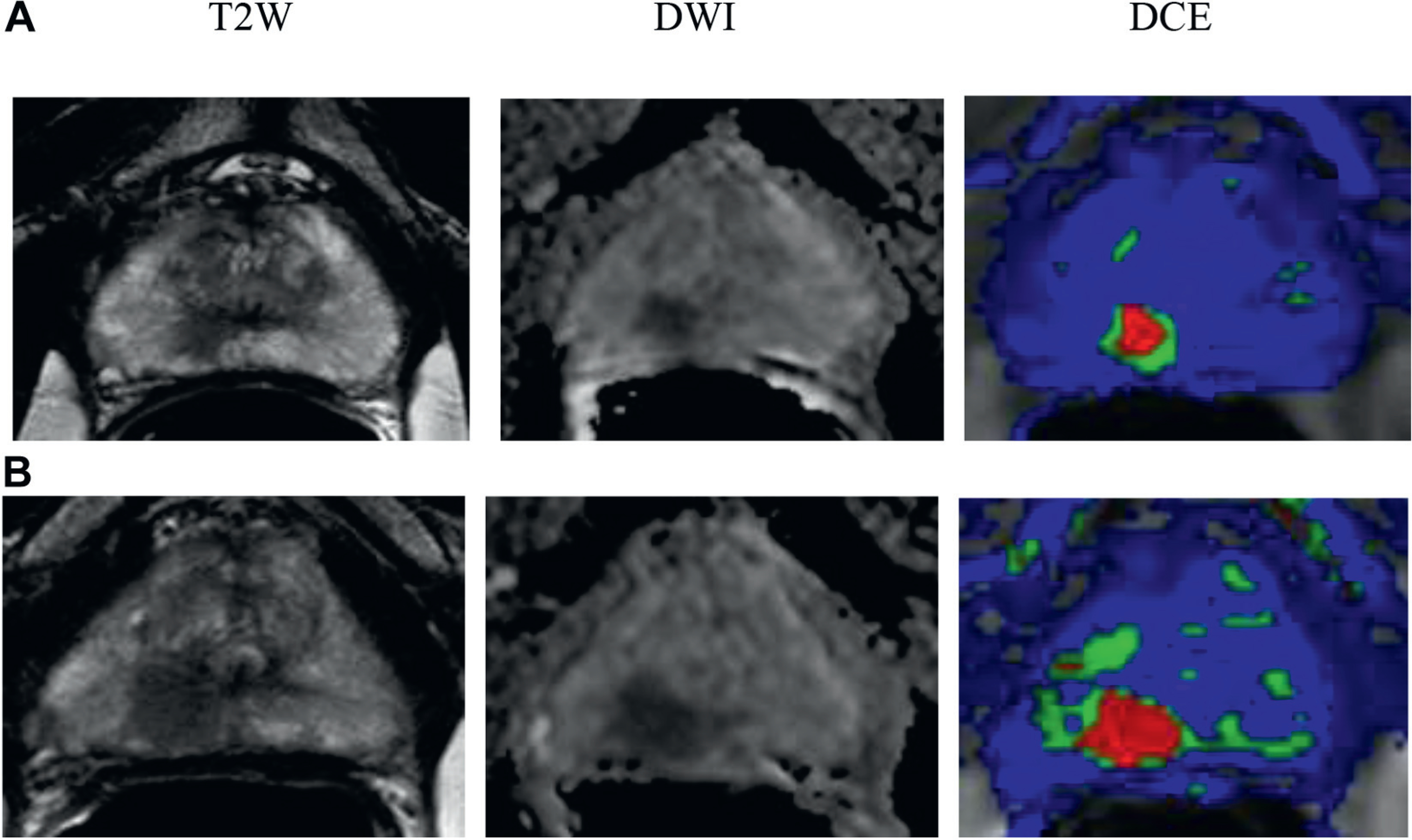
Figure 2.

In 61-year-old healthy patient with no comorbidities PSA was 3.04 ng/ml. Initial MP-MRI showed 32 cc prostate and moderate suspicion for prostate cancer with 1 cm lesion in peripheral zone right apex (A). Gleason 3 + 4 = 7 was found by 2 targeted biopsy cores but all systematic biopsies were negative. Followup MP-MRI performed 18 month later showed lesion progression to 1.3 cm (B). MRI was highly suspicious for prostate cancer. Targeted biopsy revealed Gleason 4 + 4 = 8. Radical prostatectomy was done and final pathology findings were Gleason 4 + 4 = 8 and organ confined with negative margins. T2W, T2-weighted. DWI, diffusion-weighted image. DCE, dynamic contrast enhanced.
