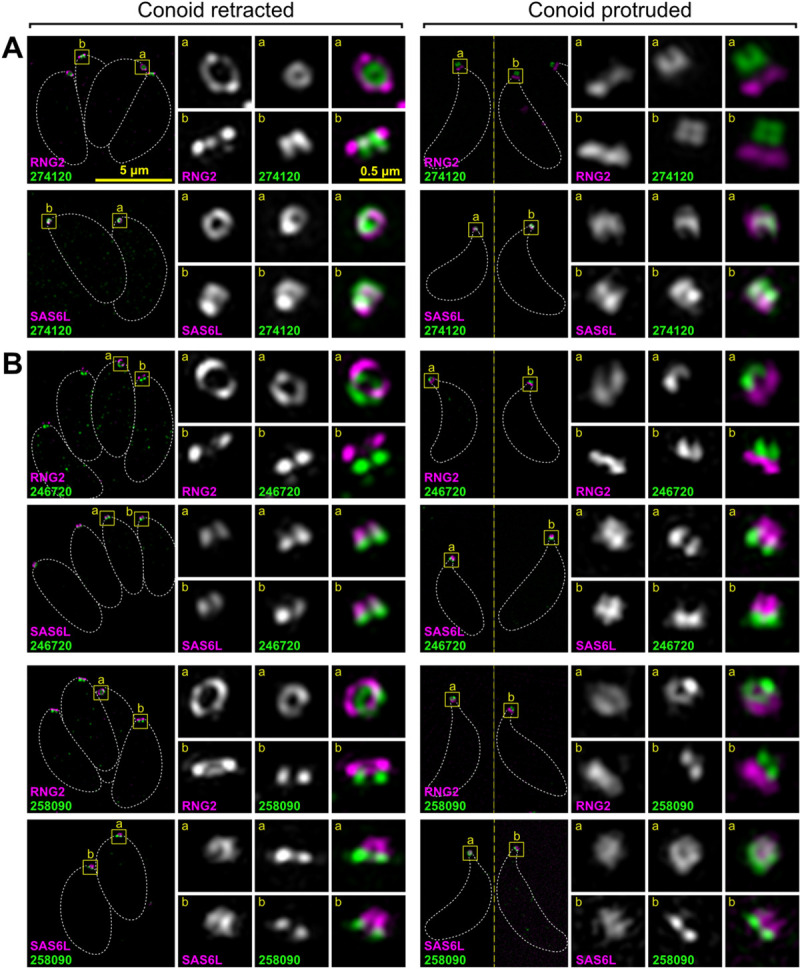
Fig 3. Super-resolution imaging of T. gondii proteins at the conoid body and base.

Immunodetection of HA-tagged conoid proteins (green) in cells coexpressing either APR marker RNG2 or conoid marker SAS6L (magenta) imaged either with conoids retracted with parasites within the host cell, or with conoids protruded in extracellular parasites. (A) Example of protein specific to the conoid body and (B) examples of proteins specific to the conoid base. See S2 and S3 Figs for further examples. All panels are at the same scale, scale bar = 5 μm, with zoomed inset from white boxed regions (inset scale bar = 0.5 μm). Dashed white lines indicate the cell boundary. APR, apical polar ring; HA, hemagglutinin; SAS6L, SAS6-like.
