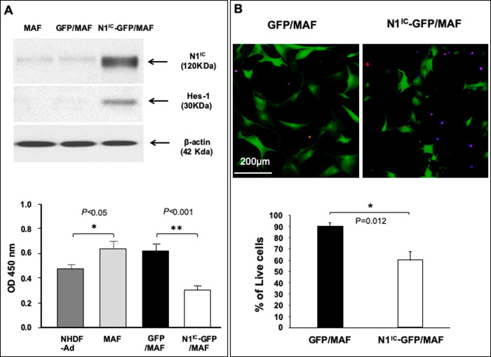
Fig 3. The Notch1 activation decrease MAF cell viability and proliferation.
A. Top: overexpression of N1IC in N1IC-GFP/MAF detected by immunoblot. increased Hes-1 expression demonstrates that the Notch pathway is activated. β-actin was used as loading control. Bottom: high intracellular Notch1 activity inhibits MAF proliferation in vitro as determined by WST cell proliferation assay. B. High activity of the intracellular Notch1 pathway decreases MAF cell viability. Top: images of cells in co-staining assay. Dead cells were double-positive [TUNEL (Red)+ and active caspase-3 (Blue)+], indicating that they are apoptotic cells. A single scale bar in a panel of pictures is representative for all pictures. Bottom: % of live cells. All data are presented as mean ± SD based on three independent experiments.

