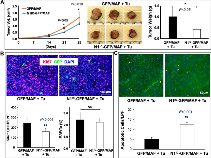
Fig 5. Increasing Notch1 activity in MAFs inhibits melanoma growth in vivo.
A. left: tumor growth curve. middle: three representative images of resected tumors from each group [Tu stands for tumor cells (1205Lu)]. right: Tumor weight measured right after resection 4 weeks post co-grafting (n = 6 tumors/group). B. Immunostaining of tumor tissues. Fewer Ki67+ cells are detectable in 1205Lu co-grafted with N1IC-GFP/MAF compared to that co-grafted with GFP/MAF. Top: representative images; Bottom: number of Ki67+ cells per low power field (LPF, 10X) and % of N1IC-GFP/MAF vs. GFP/MAF in resected tumor tissues per LPF. C. More apoptotic tumor cells were detectable in 1205Lu co-grafted with N1IC-GFP/MAF vs. GFP/MAF. Top: representative images. Apoptotic tumor cells were co-stained with TUNEL (Red) and Luc (Green) and double-positive cells (orange color); Bottom: number of apoptotic cells per low power field (LPF, 10X). All data are mean ± SD based on 5 random sections per tumor tissue, n = 6 tumors/group.

