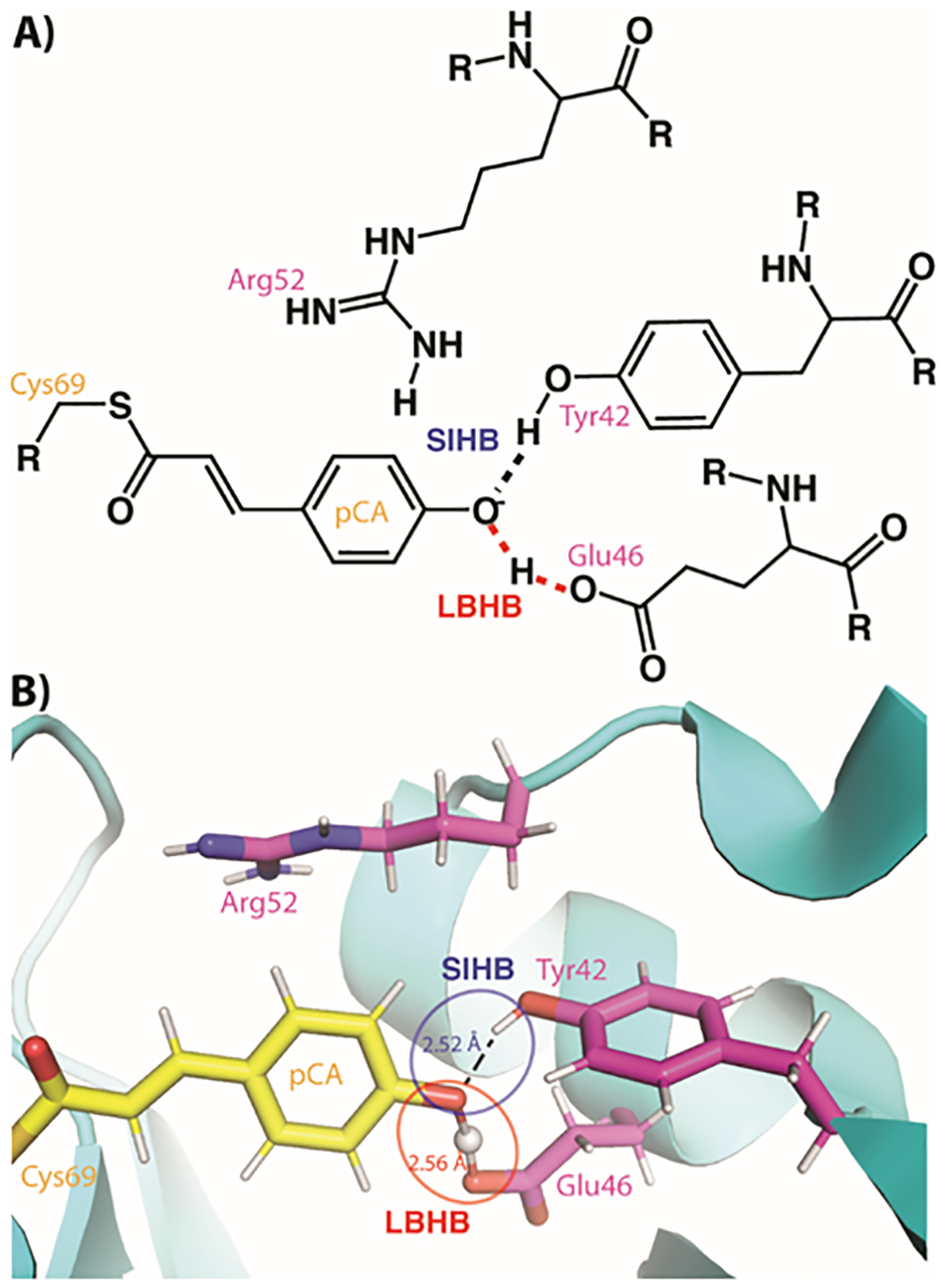
Figure 3. The chromophore binding site in photoactive yellow protein (PYP).

A) A schematic of the PYP chromophore (pCA) binding site with the two types of SHBs indicated. R groups indicate continuation of the protein chain. B) The neutron crystal structure of the PYP chromophore active site with the two types of SHBs indicated.
