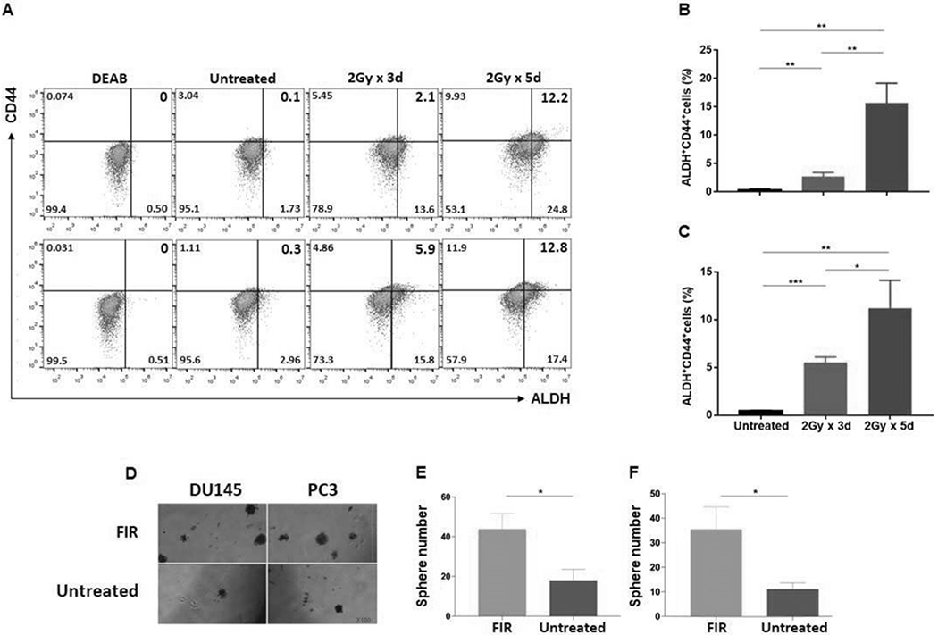
Figure 1. PCSCs are FIR-resistant.

PCa cells were irradiated at doses and times as indicated, and after 24hrs analyzed for ALDH+CD44+ cells by flow cytometry. Data are shown for DU145 (A: top) and PC3 (A: bottom) cell lines. ALDH+CD44+ PCSCs (%) are presented in the upper right quadrant. DEAB, an inhibitor of ALDH1/3 isoforms, was used to establish the baseline fluorescence as the gating reference standard of ALDH- population. Mean± SD of ALDH+CD44+ cells (%) are shown for cell lines DU145 (B) and PC3 (C). Cells plated in 6-well cell culture plates (Corning) at a density of 5×10⁵ cells/well in 2 mL RPMI 1640 medium containing 10% FBS were treated with FIR (2Gy/day x 5 days). Cells were trypan blue stained to exclude dead cells and living cells were tested for their ability to form spheres. After 7-10 days, small spheres were detected by microscopy. At day 18, spheres/well were counted. Representative photographs of DU145 and PC3 cells lines are shown (100x) (D) as well as mean ± SD of spheres/well for DU145 (E) and PC3 (F). The experiments were repeated 3 times. *p<0.05; **<0.01; ***<0.001.
