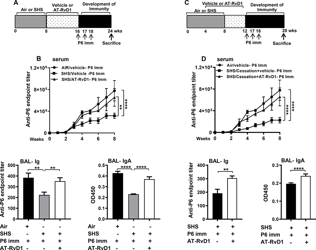
FIGURE 5. AT-RvD1 treatment of SHS-exposed mice augments the efficacy of P6 vaccination.
(A) Mice exposed to air or SHS for 8 wks and then treated with AT-RvD1 or vehicle for an additional 8 wks were vaccinated with purified P6 lipoprotein as described in methods. (B) Total P6-specific IgG Ab titers were quantified by ELISA. in weekly serum (upper panel) collected after the start of vaccination and in end-point BAL (lower left panel) collected at the time of euthanasia Levels of mucosal anti-P6 IgA (lower right panel) were quantified by measuring OD450 values in the BAL at dilutions of 1:400 in ELISA assays as described in methods. (C) Mice were exposed to air or SHS for a total of 8 wks, with or without AT-RvD1 treatment between wks 4–12, and then vaccinated with NTHI P6 antigen as described in methods. (D) Total P6-specific IgG Ab titers were measured by ELISA in serum (upper panel) collected weekly after P6 vaccination and in end-point BAL (lower left panel). Mucosal anti-P6 specific IgA levels (lower right panel) were determined by measuring OD450 values in the BAL at 1:400 dilutions by ELISA as described in methods. All treatment groups were performed at the same time, and data represent results generated from a single experiment using a total of n=10 mice/group at each time point. The results are depicted as mean ± SE. Statistical significance was determined either by two-way ANOVA with Tukey’s posttest for multiple comparisons (B & D) or two-tailed unpaired students t test (**p<0.01, ****p<0.0001).

