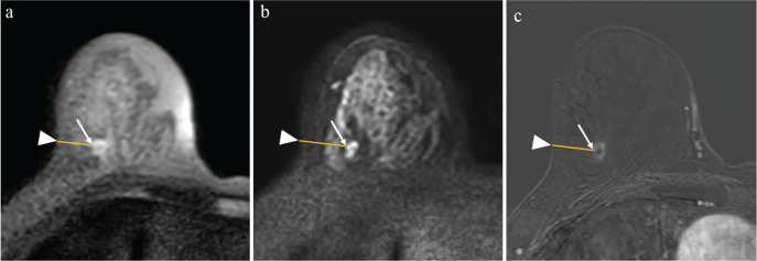
Fig. 2.
A 60-year-old woman diagnosed with right-invasive ductal carcinoma by lumpectomy. The methods used to evaluate geometric distortion in TSE-DWI and EPI-DWI. Two matching anatomic sites were used: the site of the lesion closest to the skin (arrows), and the site of the skin closest to the lesion (arrowheads). The distance between the sites was calculated as DistanceTSE in images of TSE-DWI (b = 850 s/mm2) (a), DistanceEPI in those of EPI-DWI (b = 850 s/mm2) (b), and DistanceDCE-MRI in those of DCE-MR (c). In this case, DistanceTSE, DistanceEPI, and DistanceDCE-MRI were 19.1, 25.3, and 19.5 mm, respectively. The geometric distortions of TSE-DWI and EPI-DWI were 0.021 and 0.29, respectively. TSE, turbo spin-echo; DWI, diffusion-weighted imaging; EPI, echo-planar imaging.

