Figure 1.
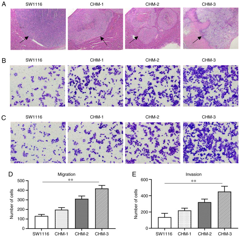
Metastatic potential is increased in the CHM-1, CHM-2 and CHM-3 cells compared with parental SW1116 cells in vivo and in vitro. (A) Hematoxylin and eosin staining of liver metastatic lesions (indicated by black arrows) formed from SW1116, CHM-1, CHM-2 and CHM-3 cells in vivo. Transwell (B and D) migration and (C and E) invasion assays were used to analyze the migration and invasion of SW1116, CHM-1, CHM-2 and CHM-3 cells. Data are presented as the means ± SD; n=3; **P<0.01.

