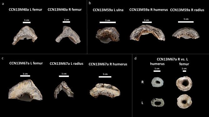
Figure 4.
Cross section of microscopic samples from Con Co Ngua. (a) Femora sections from CCN13M40a. There is increased cortical thickness, and the cortical width of right femur is asymmetrically wider than the left. (b) Upper limb sections from CCN13M40a. Large endocortical pores are evident. Medullary canal widening is most distinct on the humerus. (c) Upper and lower limb sections from CCN13M67a. While the femur presents with extreme cortical thickness and restriction of the medullary canal area, the upper limb sections present with severe porosity of the endocortical surfaces. (d) Asymmetry of the medullary canals of the humeri and femora of CCN13M67a.

