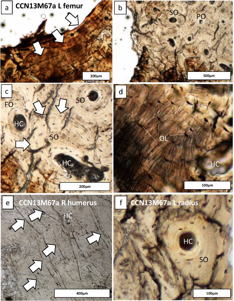
Figure 6.
Regions of bone captured from the endocortical surface in the adolescent (CCN13M67a), allowing to examine the degree of bone modelling and remodeling. (a–d) The femur: Secondary osteons (SO), primary osteons (PO), Haversian canals (HC), and osteocyte lacunae (OL) can be seen. (a) White arrows point to endosteal lamellar layers which are typical for this bone region. (c) White arrows point to a cement line of a secondary osteon that indicates a remodeling event of a fragmentary osteon (FO) underneath, confirming cyclical replacement of old bone with new bone tissue, possibly driven by increased metabolic bone needs. (e) White arrows point to primary lamellar bone layers and an isolated HC. (f) A SO amongst primary bone.

