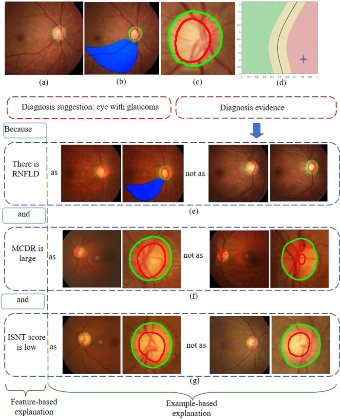
Fig. 2. Schematic of transparency and interpretability for hierarchical deep learning system.
a Original fundus image. b Segmentation of OD, OC, and RNFLD. c Magnification of the image shown in (b). d Two-dimensional plane of MCDR and ISNT score. e–g Comparison of the fundus image currently being evaluated with fundus images that have been clearly diagnosed in the database in terms of MCDR, ISNT score, and RNFLD. OD optic disc, OC optic cup, RNFLD retinal nerve fiber layer defects, MCDR mean cup-to-disk ratio, ISNT inferior, superior, nasal, temporal.

