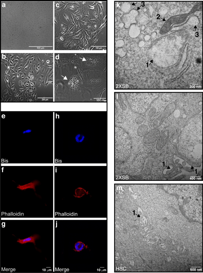
Figure 2.
The morphology of the 2XSB sporadic MPNST cell line recapitulates the pattern seen in the parent tumor. (a–d) Representative brightfield (a) and phase contrast (b–d) images of logarithmic phase 2XSB cells. Both brightfield and phase contrast microscopy show that the tumor cells have a cobblestoned to spindled morphology. Note also that the ×60 phase contrast images in (c) show several cytoplasmic vacuoles in the tumor cells, while (d) illustrates two representatives of the subpopulation of large multinucleated cells present in this line (arrows). (e–j) Two representative 3D culture images (g,j) show the merged nuclei and cytoskeleton images acquired at ×63 with z-stack microscopy highlighting spindled (e–g) and polygonal (h–j) multinucleated cells. Panels (e–i) show the individual channels acquired for phallodin-Texas Red (cytoskeleton labeling, f,i) and DNA-bisbenzamide (nucleus labeling, e,h) prior to merging in panels (g) and (j). (k–m) Transmission electron microscopy (k) demonstrates that 2XSB cells contain swollen (1) and normal (2) mitochondria and swollen rough endoplasmic reticulum (3). Both 2XSB cells and non-neoplastic human Schwann cells also have numerous microvilli on their cellular surfaces (1 in l,m).

