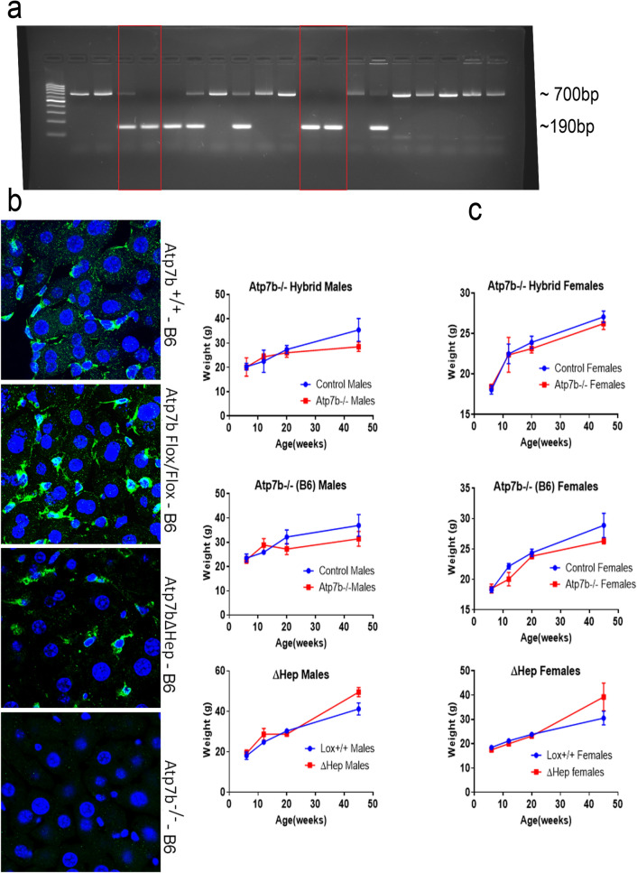
Figure 1.
Characteristics of Atp7b−/− mice on C57BL/6 J background (Atp7b-/–B6). (a) Genotyping littermates to identify the Cre-mediated deletion. The 190 bp product is indicative of the desired deletion within Atp7b (red boxes). The 700 bp product reflects the wild-type genotype. (b) The livers from 6-weeks old mice were sectioned, immuno-stained with anti-ATP7B antibody (green) and DAPI (nuclei, blue), and then imaged using an LSM 800 confocal microscope. In the wild-type (Atp7b+/+) and Atp7bFlox/Flox livers, ATP7B (green) is detected in both hepatocytes (large cells with large nuclei) and non-parenchymal cells (small cells with small nuclei). Atp7bΔHep-B6 mice show staining predominantly in small non-parenchymal cells. No ATP7B staining is seen in Atp7b-/–B6 livers. (c) Comparison of the weight gain curves for Atp7b-mutant mice (blue), males and females, and the background-matched control animals (red); each point represents an average of data from 3–11animals. Individual weights and statistical analysis of weight differences between the Atp7b−/− mice and the background-matched wt controls at 20 weeks and at 45 weeks are given in Supl. Fig. S1.

