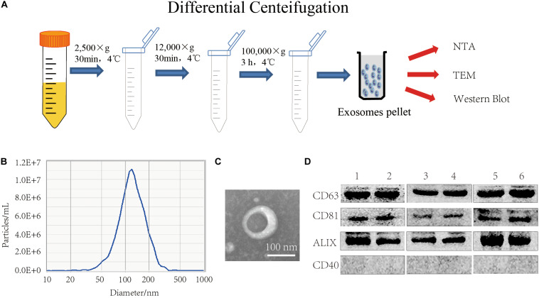
FIGURE 2.
FBS EXO extraction and identification. (A) Centrifugation; (B) NTA detection of the median particle size of FBS EXO; (C) Observation of FBS EXO structure under TEM (×100,000); (D) Western Blotting was used to detect the protein expression of exosome markers CD63, CD81, and ALIX and the microcapsule surface marker CD40 in different batches of FBS EXO, 1-6 represented exosomes extracted from different batches.

