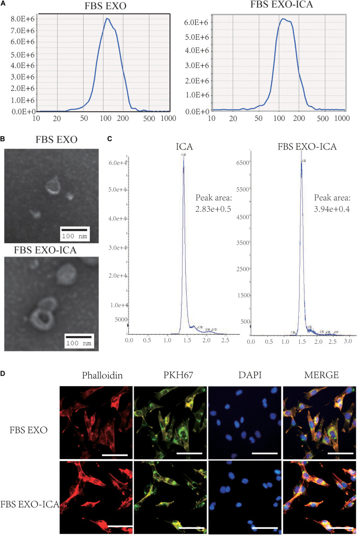
FIGURE 4.
Construction and identification of FBS EXO-ICA. (A) Particle sizes of FBS EXO and FBS EXO-ICA detected by NTA; (B) TEM results showing the typical lipid bilayer membrane structure and diameter of FBS EXO-ICA (×8,000); (C) HPLC indicating the extent of incorporation of ICA in FBS EXO; (D) Fluorescence staining showing that phalloidin labeled the cytoskeleton red, PKH67 labeled the exosome membranes green, and DAPI labeled the cell nuclei blue (×200).

