FIGURE 1.
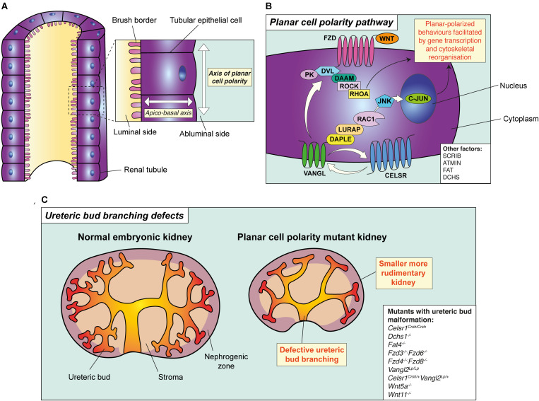
The principle and pathway of planar cell polarity and its role in branching morphogenesis. (A) The principle of planar cell polarity (PCP). An individual tubular epithelial cell is shown from a tubule cut lengthways. The axis formed between the luminal side of the cell and the abluminal side is termed the apico-basal axis. Conversely, the axis of orientation within the plane of the tubule is the axis of PCP. A key feature of PCP is the asymmetric expression of core PCP proteins, such as VANGL and FZD, setting the direction of polarity across the plane of the tubule. (B) The planar cell polarity pathway and its proteins, as demonstrated in a tubular epithelial cell. Binding of WNT ligands (WNT5A or WNT11) to FZD receptors leads to the recruitment of DVL proteins to the membrane resulting in the formation of a multi-protein complex. This complex interacts with multiple effector molecules. Downstream of DVL, two independent and parallel pathways have been proposed. The first pathway signals to RHOA through the formin homology protein; DAAM1, and ROCK. The second pathway involves the adaptor proteins DAPLE and LURAP, ultimately resulting in C-JUN-dependent transcription and coordinating planar-polarised behaviours. Other factors involved in PCP are shown in the white box. (C) PCP and ureteric bud branching. Mutation or deletion of core PCP proteins in mouse lead to the simplification of ureteric bud branching, particularly in the caudal component of the organ, accompanied by a reduction in kidney size. All mouse models with disruption of PCP proteins which have also been demonstrated to have defective ureteric bud branching defects are shown in the white box.

