Fig. 5.
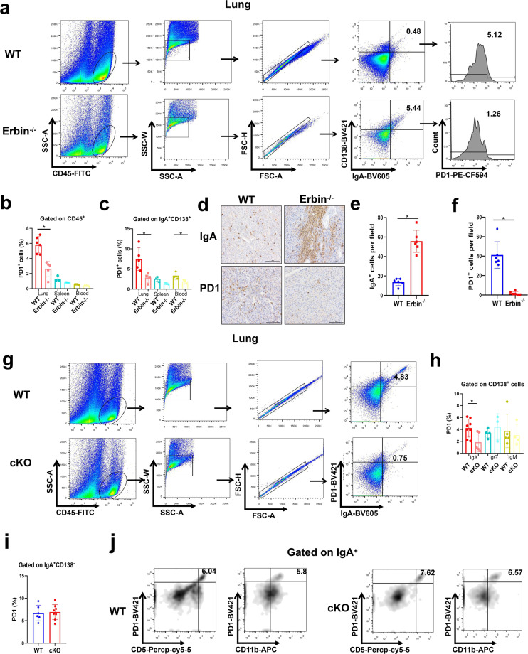
PD1 expression in IgA+ CD138+ cells in lung metastases of CRC in Erbin-deficient mice. a The flow cytometry analysis of PD1+ cells gated on IgA+ CD138+ cells in lung of tumor-bearing WT and Erbin−/− mice. b The percentage of PD1+ cells in leukocytes of lung, spleen, and blood from tumor-bearing WT and Erbin−/− mice. c The percentage of PD1+ cells gated on IgA+ CD138+ cells in lung, spleen, and blood of tumor-bearing WT and Erbin−/− mice. d–f Immunohistochemical staining of IgA and PD1 and semi-quantification of the number of IgA+ (e) and PD1+ cells (f) in lung metastases from tumor-bearing WT and Erbin−/− mice. Scale bar, 100 μm. g–j The flow cytometry analysis of PD1+ IgA+ cells gated (g), the percentage of PD1+ cells gated on IgA+/IgG+/IgM+ CD138+ cells (h), the percentage of PD1+ cells gated on IgA+ CD138− cells (i), the flow cytometry analysis of CD5+/CD11b+ PD1+ cells gated on IgA+ cells (j), in lung leukocytes of tumor-bearing WT and cKO mice. *p < 0.05 (Student t test)

