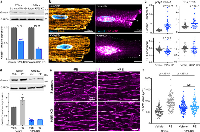
Fig. 6. mRNA localization and cardiac hypertrophy depends on the microtubule motor Kinesin-1.
a (top) Representative western blot images of kinesin-1 and loading control GADPH in ARVMs. (bottom) Quantification of relative kinesin-1 expression, normalized to GAPDH. N = 3. b Representative immunofluorescence images of the microtubule network (left) and polyA mRNA (right) in Scramble and Kif5b KD cells. White arrow indicates ICD. c Nuclear:cytosolic and ICD:cytosolic ratios of polyA mean fluorescence (left) and 18s rRNA mean fluorescence (right) in Scramble and Kif5b KD cells. Scramble (N = 2, n = 62, polyA) (N = 2, n = 88, 18 s), Kif5b KD (N = 2, n = 61, polyA) (n = 2 , N = 76, 18 s). Statistical significance determined via two sample, two-tailed t-test. d (top) Representative western blot images of kinesin-1 and loading control GADPH in NRVMs. (bottom) Quantification of relative kinesin-1 expression, normalized to GAPDH. N = 3. e Representative live-cell images of (top row) Scramble or (bottom row) Kif5b KD NRVMS, treated with (left column) vehicle or (right column) PE. f Quantification of cell areas in NRVMs from experiment shown in e. Scramble+vehicle (N = 3, n = 160), Scramble+PE (N = 3, n = 160), Kif5b KD + vehicle (N = 3, n = 160), Kif5b KD + PE (N = 3, n = 160). For all graphs shown in figure, the mean line is shown, with whiskers denoting standard error (SE) from the mean. Source data are provided as a Source data file.

