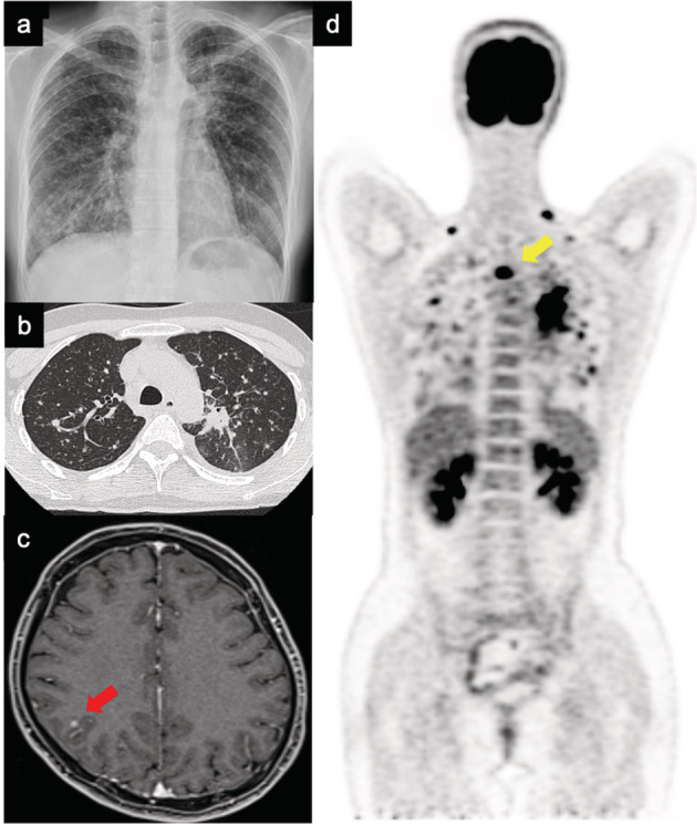
FIGURE 1.

Images at the time of lung cancer diagnosis. (a) Chest X‐ray and (b) chest computed tomography showing primary lesion in the upper left lobe and multiple lung metastases. (c) Brain magnetic resonance image. The red arrow points to the brain metastasis. (d) Whole‐body scan using positron emission tomography. The yellow arrow points to the bone metastatic lesion
