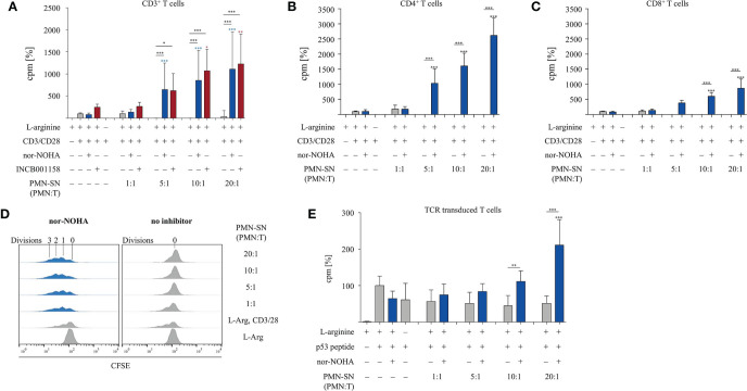
Figure 1.
T cell proliferation is strongly enhanced by polymorphonuclear neutrophil granulocytes supernatants (PMN-SN) in the presence of arginase inhibition. (A–C) Primary human T cells (bulk CD3+ or sorted CD4+ or CD8+) and PMN were isolated from whole blood of healthy donors. PMN were pre-incubated in cell culture medium for 72 h in the presence or absence of the arginase inhibitor nor-NOHA (1 mM) or INCB001185 (100 µM). T cells were then stimulated for 48 h with anti-CD3/anti-CD28-tagged beads in the PMN-SN. T cell proliferation was determined by [3H]thymidine incorporation over 16 h and values of stimulated cells in the presence of arginine were set as 100%. (A) CD3+ T cells from n=31 individual experiments (n=5 for INCB001185) with T cells from different blood donors, (B) CD4+ T cells (n=4) and (C) CD8+ T cells (n=4). (D) Human PBMC and PMN were isolated from whole blood of healthy donors. T cells were retrovirally transduced with a HLA-A2 restricted p53(264-272)-specific T cell receptor and expanded by weekly restimulation with anti-CD3/anti-CD28-tagged beads and later via p53 peptide-loaded K562-A2.1 cells. PMN-SN was generated as described in (A–C). T cells were then stimulated with p53 peptide-loaded irradiated K562-A2.1 cells in PMN-SN for 48 h. T cell proliferation (n=3) was analyzed as in (A–C). (E) Primary human CD3+ T cells were activated in PMN-SN for 48 h as described in (A). T cell proliferation was determined by carboxyfluorescein succinimidyl ester (CFSE) staining (one representative experiment of n=7 is shown). Unless otherwise stated, statistical analysis refers to the control conditions of activated T cells in the presence of L-arginine and nor-NOHA. Statistical calculations were performed with one-way ANOVA and Tukey´s post hoc test (***p < 0.001, **p < 0.01, *p < 0.05).

