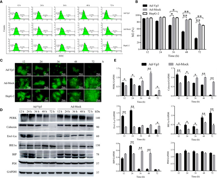
Figure 2.
Endoplasmic reticulum related detection. The endoplasmic reticulum stress of Ad-Vp3-infected HepG-2 cells can be stimulated by Apoptin, 24 h and 48 h post-infection, and ER functional structure is gradually impaired gradually from 36 h to 72 h post-infection. (A, B) Flow cytometry detection of ER using the ER-Tracker™ Green. (C) Endoplasmic reticulum ER-Tracker™ Green staining (200×). (D, E) Western-blot detection of endoplasmic reticulum stress related proteins. Data are presented as mean ± SD, *p < 0.05, **p < 0.01.

