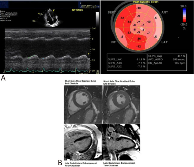
Figure 2.

(A) Transthoracic echocardiography was performed on Day 3 of admission whilst the patient was on dobutamine infusion. This demonstrated a tricuspid annular plane systolic excursion of 11.7 mm and an average left ventricular global longitudinal strain of -8.7%. (B) Cardiac magnetic resonance imaging performed on Day 8 of admission after clinical recovery. Short axis cine gradient echo still (end diastole and end systole) images of the heart at mid cavity level showed normal biventricular contraction. Long axis views of the heart demonstrated no late gadolinium enhancement. There was no imaging evidence of myocardial infarction or fibrosis.
