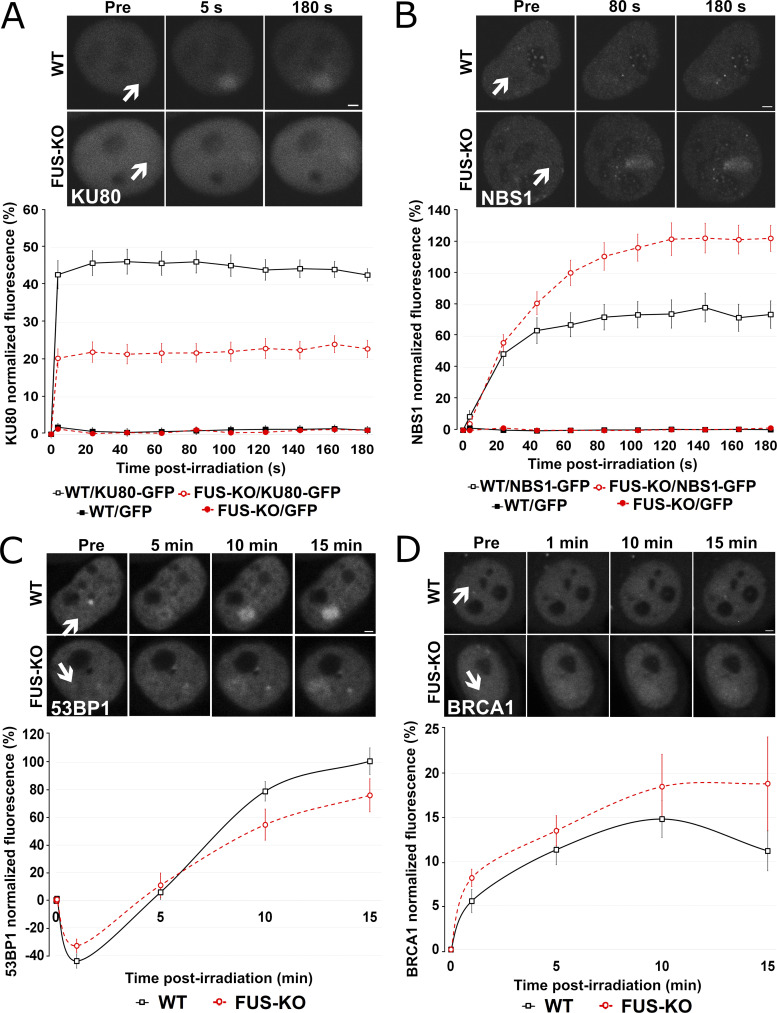
Figure 3.
Loss of FUS changes the pattern of recruitment of HR- and NHEJ-related proteins to DSBs. (A) HeLa WT and FUS-KO cells were transiently transfected with a KU80-GFP expressing plasmid. Upper panel: Representative micrographs of selected time points. Lower panel: Time course of the normalized fluorescence intensity of KU80-GFP recruitment at the microirradiated sites. All microirradiation experiments were performed in two biological replicates (with 10 cells each, except for BRCA1 recruitment, which was done in four biological replicates with 10 cells each). (B) HeLa WT and FUS-KO cells were transiently transfected with a NBS1-GFP–expressing plasmid. Upper panel: Representative micrographs of selected time points. Lower panel: Time course for NBS1-GFP recruitment. (C) HeLa WT and FUS-KO cells were transiently transfected with a 53BP1-GFP–expressing plasmid. Upper panel: Representative micrographs of selected time points. Lower panel: Time course for 53BP1-GFP recruitment. (D) HeLa WT and FUS-KO cells were transiently transfected with BRCA1-GFP–expressing plasmids. Upper panel: Representative micrographs of selected time points. Lower panel: Time course for BRCA1-GFP recruitment. In all graphs, data are plotted as normalized average ± SEM. Scale bars: 2 µm. Arrows indicate microirradiated area.

