Figure 4.
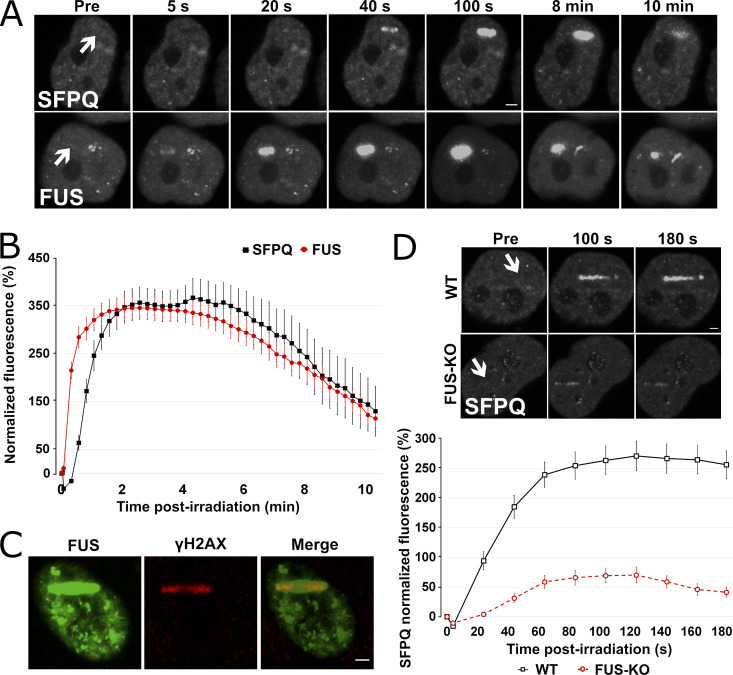
FUS recruitment to DSBs precedes SFPQ, and its absence strongly reduces SFPQ accumulation. (A) WT HeLa cells were transiently transfected either with SFPQ-GFP or FUS-GFP plasmid and then laser microirradiated. (B) Comparison of FUS and SFPQ recruitment kinetics. (C) HeLa cells were transiently transfected with a GFP-tagged FUS expression plasmid and then laser microirradiated. Cells were then immunostained for γH2AX. H2AX is phosphorylated at laser microirradiation sites and colocalized with FUS-GFP. (D) HeLa WT and FUS-KO cells were transiently transfected with a SFPQ-GFP–expressing plasmid. Upper panel: Representative micrographs of selected time points (see Video 1). Lower panel: Time course for SFPQ-GFP recruitment. In all graphs, data are plotted as normalized average ± SEM. Scale bars: 2 µm. Arrows indicate microirradiated area.

