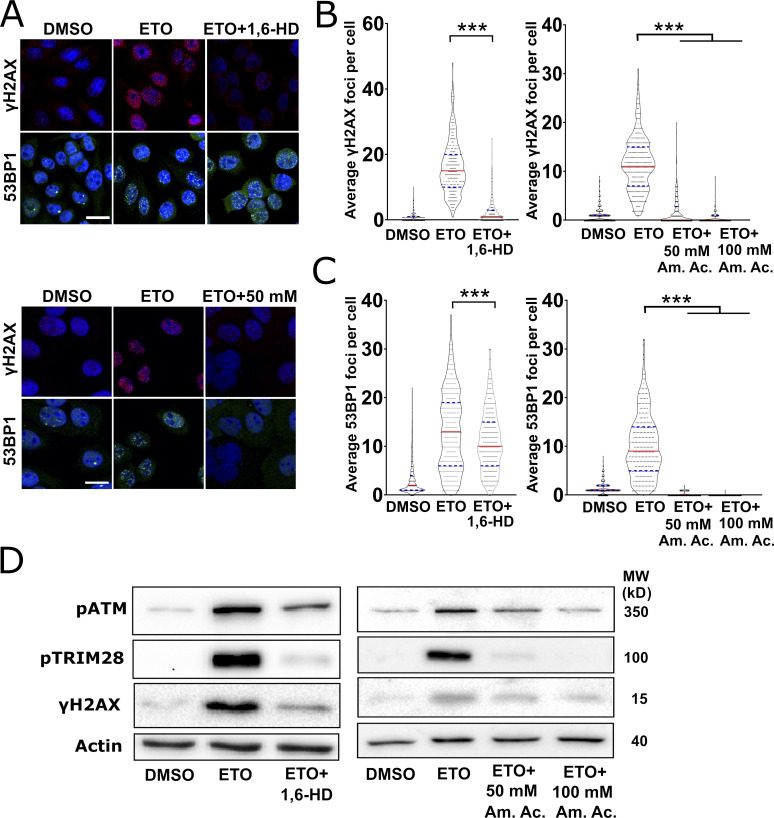
Figure 7.
LLPS is required for DDR activation and foci formation. (A) Representative confocal micrographs of HeLa cells that were treated with ETO alone, ETO plus 1,6-HD (upper micrographs), or ETO plus 50 or 100 mM Am. Ac. (lower micrographs) before immunostaining for γH2AX or 53BP1. Scale bars: 20 µm. (B) Quantification of γH2AX 1 foci in the experiments in A. In the HD experiment, 200 cells were analyzed per condition, and in the Am. Ac. experiment, 150 cells per condition, and experiments were performed in duplicate. Graphs represent the number of foci per cell and are shown as violin plots with all samples. Statistics: one-way ANOVA and Bonferroni post hoc test. (C) Quantification of 53BP1 foci in the experiments in A. Experiments, quantifications, and statistics were performed as described in B. (D) Western blot analysis of total extracts prepared from HeLa cells treated with ETO alone, ETO and 2% 1,6-HD, or ETO and Am. Ac. (50 or 100 µM). Phosphorylation of ATM, TRIM28, and H2AX was assessed (loading control: Actin). MW, molecular weight. ***, P < 0.001.

