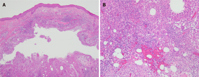
Figure 2.
Pathological biopsy of the lesion was performed. A: A cystic cavity can be seen in the dermis, surrounded by granulomatous structures formed by histiocytes [Hematoxylin-eosin staining (HE) × 40]; B: Scattered around the cystic cavity are multinucleate giant cells, accompanied by lymphocyte, plasma cell, and neutrophil infiltration (HE × 200).

