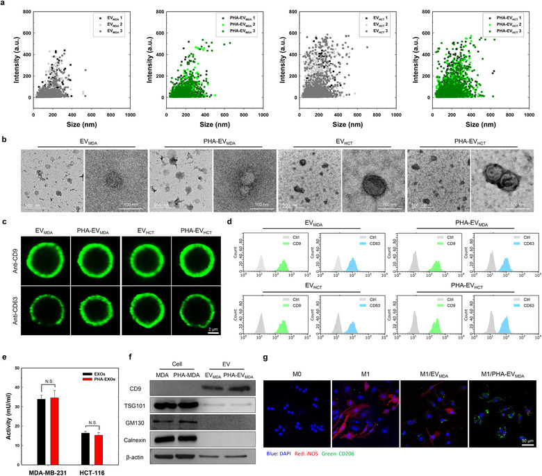
FIGURE 4.

Physicochemical characteristics of PHA‐EVs. (a) Size distribution of bare EVs and PHA‐EVs (n = 3). (b) TEM images of bare EVs and PHA‐EVs. (c) Confocal microscopy images of surface markers on bare EVs and PHA‐EVs. (d) Flow cytometry analysis of surface markers on bare EVs and PHA‐EVs. (e) AchE activity of bare EVs and PHA‐EVs (n = 3). (f) Western blotting analysis of bare EV‐secreting cells, PHA‐EV‐secreting cells, bare EVs and PHA‐EVs. (g) M1–M2 macrophage polarization by the PHA‐EVs. Confocal microscopy images show iNOS (red) and CD206 (green) in cells
