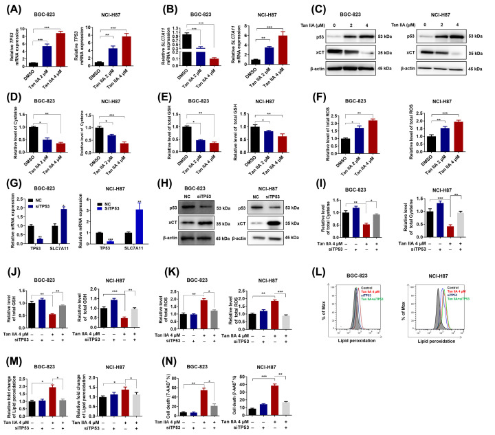
Figure 3. p53 mediates Tan IIA-induced ferroptosis in BGC-823 and NCI-H87 gastric cancer cells.
(A,B) BGC-823 and NCI-H87 cells were treated with 2 and 4 μM Tan IIA for 72 h, the mRNA expression of TP53 and its target gene SLC7A11 was detected by RT-qPCR. (C) BGC-823 and NCI-H87 cells were treated with Tan IIA as mentioned above. The protein expression of p53 and xCT was detected by Western Blot. (D–F) BGC-823 and NCI-H87 cells were treated with Tan IIA as mentioned above. Intracellular cysteine level, GSH level and ROS level were measured. (G,H) BGC-823 and NCI-H87 cells were transfected with 20 μM TP53 siRNA and negative control (NC) sequence using Lipofectamine 3000 for 4 h, after 24 h more culture with fresh RPMI-1640 medium with 10% FBS, cells were harvested. The knockdown efficiency of TP53 and the expression of its target gene SLC7A11 in both BGC-823 and NCI-H87 gastric cancer cells was detected by RT-qPCR and Western Blot. (I–K) BGC-823 and NCI-H87 cells were pretreated with TP53 siRNA, then the cells were treated with Tan IIA for 72 h, intracellular cysteine level, GSH level and ROS level were measured. (L–N) BGC-823 and NCI-H87 cells were treated with TP53 siRNA and Tan IIA as mentioned above. Then lipid peroxidation and cell death was detected by flow cytometry. Data are shown as the mean ± SD (n=3). *P<0.05 vs control group; **P<0.01 vs control group; ***P<0.001 vs control group.

