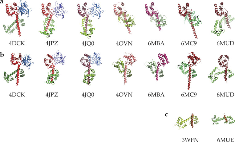
Figure 2.
Published crystal structures of CTNaV with CaM. (a) Structures aligned pairwise to CTNaV1.5–CaM (PDB accession no. 4OVN) using the EFL domain as an anchor. Each CT is in shades of red, CaM in green, and FHF in blue. The angle of helix αVI with respect to the EFL varies in these constructs. (b) The same structures aligned pairwise using the CaM C-lobe as an anchor and so helix αVI has the same orientation. These alignments highlight the different relative orientations of the EFL and helix αVI, displaying the EFL to the right (4OVN, 6MBA, 6MC9) or to the left (4DCK, 4JPZ, 4JQ0, 6MUD). (c) Complexes of CaM with shorter peptides of CTNaV1.5.

