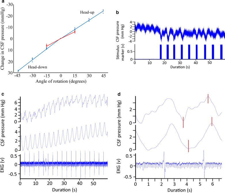
Fig. 5.
a Data from the cranial (blue) and spinal (red) implantation sites during rotations. Separate curve fitting was done for the head-up and head-down rotations, though the slopes were not significantly different. Rotations resulted in significantly lower changes in CSF pressure at the spinal recording site (red) compared to the cranial recording site (blue). b Raw simultaneous data recordings of CSF pressure and electrical stimulation of the suboccipital muscle in Alligator mississippiensis. Stimulation of the muscle was associated with a decrease in CSF pressure. Variation in this response may represent variation in stimulus strength (caused by slight displacement of the muscle relative to the stimulating probe) or possibly fatigue of the muscle. c Simultaneous recordings of CSF pressure from the cranium (top) and spinal canal (middle) of A. mississippiensis. These two CSF traces show clear synchrony, and are synchronized with the (simultaneously recorded) EKG trace (bottom). d Simultaneous recordings of EKG (bottom trace) as well as CSF pressure from the cranium (top) and spinal canal (middle) of A. mississippiensis. There is a temporal offset between the cranial and spinal CSF pulses (red vertical lines), such that the spinal pulse lags behind the cranial pulse by approximately 200 ms

