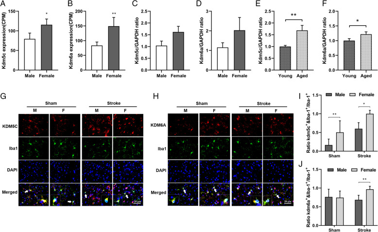
Fig. 1.
KDM5C and KDM6A expression is sexually dimorphic in microglia. a, b Flow cytometry sorted microglia were subjected to RNA sequencing, and the results showed aged (18–22 months) female microglia express higher levels of Kdm5c and Kdm6a compared to male microglia. c–d Kdm5c and kdm6a mRNA levels were measured by RT-QPCR in flow-sorted microglia from young (8–12 weeks) male and female mice, and there was no significant sex difference. e, f The mRNA levels of kdm5c and kdm6a were quantified by RT-QPCR in flow-sorted microglia cells from young and aged female mice, and the results showed aged female microglia express significantly higher levels of Kdm5c and Kdm6a compared to young female microglia. g, h 63x microscopic fields demonstrate the co-localization of KDM5C/KDM6A and Iba-1; arrows indicate cells with colocalized signals enlarged in the inserts. i, j Quantification of the ratios of KDM5C+/KDM6A+&Iba-1+ cells over total Iba-1+ cells. n=5 animals/group; *p < 0.05; **p < 0.01 female versus male mice. Scale bar = 20 μm

