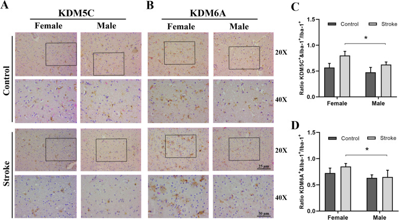
Fig. 2.
Microglia from aged female stroke patients express more KDM5C and KDM6A than male ischemic microglia. Representative immunohistochemistry microphotographs depicting KDM5C/KDM6A and Iba-1 staining of postmortem human brain tissue from acute ischemic stroke subjects and age-matched control (> 60 years). a, c 20X and 40X fields (boxed areas in 20X images) demonstrate the co-localization of KDM5C/KDM6A and Iba-1in microglia; scale bar = 25 μm and 50 μm, respectively. b, d Quantification of the ratios of KDM5C+/KDM6A+&Iba-1+ cells over total Iba-1+ cells (N = 6 control and 6 stroke subjects/group). *P<0.05

