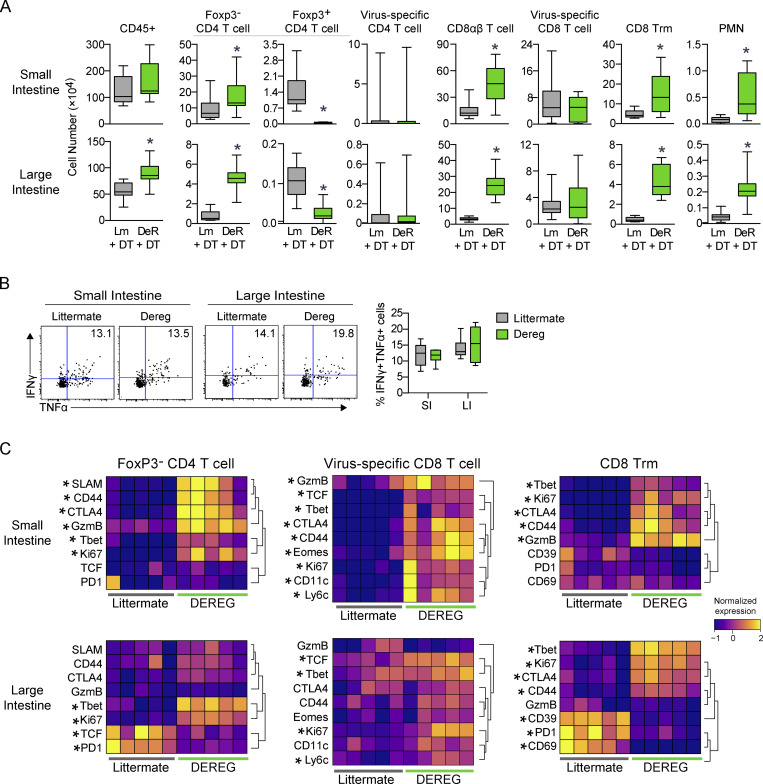
Figure 8.
Depletion of T reg cells increases virus-specific T cell activation states and increases bystander T cells in the chronically infected GIT. (A–C) DeR or Lm control mice received virus-specific CD4 SMARTA and CD8 P14 T cells and were subsequently infected with LCMV-Arm (acute) or LCMV-Cl13 (chronic). Cells from the SI and LI were analyzed by CyTOF 50 dpi. (A) The numbers of the indicated cell populations in the SI (top row) and LI (bottom row). Data from two independent experiments with n = 12 mice per group. *, P < 0.05 by t test. Error bars indicate the highest and lowest values. (B) Representative flow plots (left and center panels) and box plots (right panel) show the proportion of IFNγ+TNF+ P14 T cells following ex vivo stimulation with LCMV-GP33–41 peptide. Shown is one of three independent experiments with n = 5–6 mice per group. Error bars in box plots indicate the highest and lowest values. (C) Heatmaps show row-normalized protein expression from CyTOF analysis in the indicated immune cell populations. Each column represents an individual mouse from one of two independent experiments with n = 12 total mice per group. *, P < 0.5 (t test).

