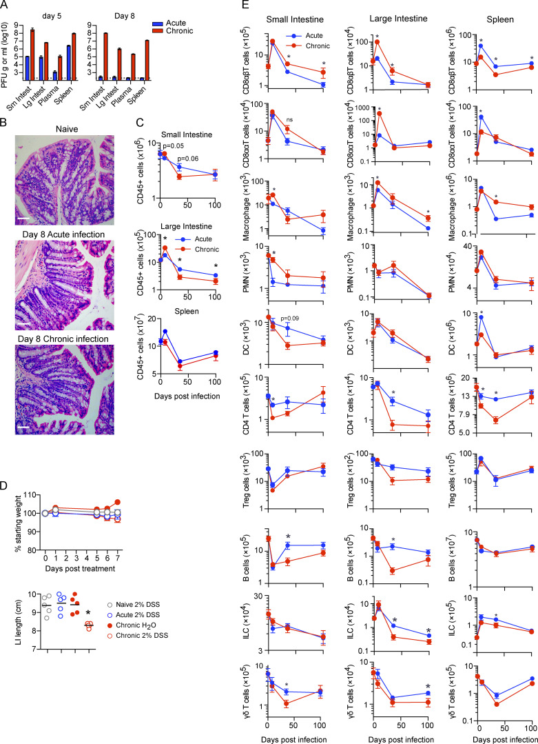
Figure S1.
The GIT is a long-lived reservoir of LCMV-Cl13 infection. (A and B) Mice were infected with LCMV-Arm (acute) or LCMV-Cl13 (chronic), and 5 and 8 dpi, PFU of virus per gram of tissue or per milliliter of blood plasma was determined. n = 4–5 mice per group from one experiment. *, P < 0.05 by ANOVA test. (B) H&E staining was performed on LI tissue from naive mice or 8 dpi after acute LCMV-Arm or chronic LCMV-Cl13 infection, showing a lack of epithelial or villus pathology during infection. Representative pictures from n = 5 mice per group from one experiment. Scale bars = 50 µm. (C and D) Mice were infected with LCMV-Arm or LCMV-Cl13, and 0, 8, 35, and 100 dpi, the SI, LI, and spleen were analyzed by CyTOF. Graphs show total number of (C) CD45+ viable cells and (D) the indicated immune cell subsets: CD8αβT cells (TCRβ+CD8α+CD8β+), CD8ααT cells (TCRβ+CD8α+CD8β−), monocytes/macrophages (CD11b+SiglecF−TCR−B220-CD11c−), polymorphonuclear cells (CD11b+Ly6G+TCR−B220−CD11c−), DCs (CD11chiMHC-IIhiTCR−B220−), CD4 T cells (TCRβ+CD4+), B cells (B220+MHC-II+), ILCs (lin−Thy1.2+), and γδT cells (TCRγδ+TCRβ−B220−) in the SI, LI, and spleen. *, P < 0.05 (Mann-Whitney test of log-transformed data). n = 10 mice per group. Error bars indicate SEM (A–D).

