Figure 4.
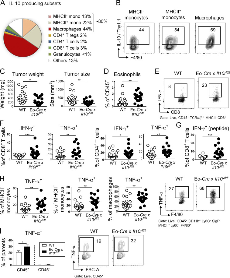
Eosinophil activities in the TME are suppressed by IL-10. (A and B) IL-10 reporter (10BiT) mice were subcutaneously injected with 5 × 10⁵ MC38 cells and analyzed after 15 d with respect to their intratumoral frequencies of Thy1.1 (IL-10)+ myeloid cells, granulocytes, and T cells. Average frequencies as assessed in independent tumors are shown in A alongside representative FACS plots for the indicated major IL-10–producing myeloid populations in B (n = 8 tumors). (C–I) Eo-Cre × Il10rafl/fl mice and their Cre-negative littermates were subcutaneously injected with 5 × 10⁵ MC38 cells and analyzed after 15 d with respect to their tumor weights and volumes (C), their intratumoral frequencies of eosinophils (D), and their intratumoral frequencies of IFN-γ+ and TNF-α+ CD4+ and CD8+ T cells (F, representative FACS plots in E; PMA and ionomycin) and IFN-γ+ CD8+ T cells upon restimulation with MC38-specific peptide (G). (H) Frequencies of TNF-α+ monocytes and macrophages among their respective parent populations, shown alongside representative FACS plots for macrophages. (I) Frequencies of TNF-α+ cells among CD45+ leukocytes and among all CD45− cells in the tumor. Data in C–I are pooled from three independent studies, n = 17–20 tumors per genotype. *, P < 0.05; **, P < 0.01; and ***, P < 0.001; as calculated by Mann–Whitney test.

