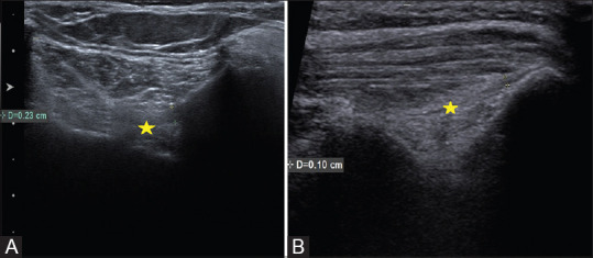
Figure 3(A and B).

(A and B): Ultrasound images of iliolumbar ligament at presentation (A) and at 6 weeks post PRP follow up (B), demonstrating regression in thickness of iliolumbar ligament from 2.3 mm to 1.0 mm. [Yellow asterisk: Iliolumbar ligament]
