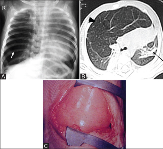
Figure 16(A-C).

Congenital lobar overinflation. (A) Hyperinflated right lung (arrow) with persistent vascular markings, distinguishing it from pneumothorax. (B) Axial CT scan of the same patient confirms overinflation (arrowhead) with mediastinal shift to the left and lingular compressive atelectasis (dotted arrow). (C) Intra-operative image with spongy appearance of lung
