Figure 6.

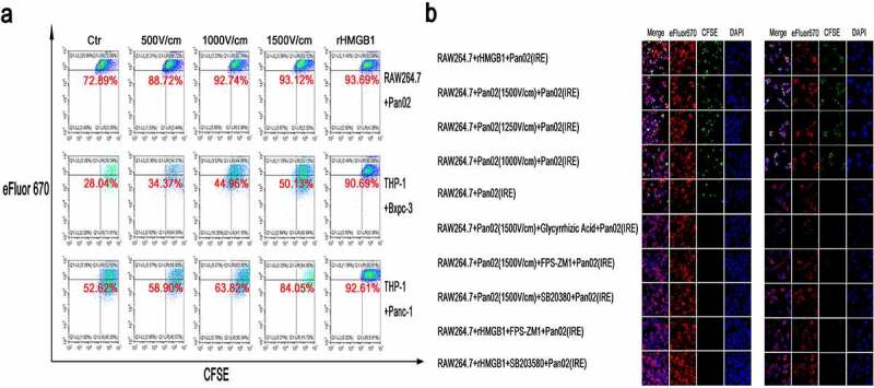
The enhancement of phagocytosis in macrophages stimulated by rHMGB1 or TSN of tumor cells treated with IRE. (a) The detection of the ability of phagocytosis of tumor cells after IRE treatment in macrophages stimulated by rHMGB1 and TSN of tumor cells treated with electric fields. Upon the stimulation of rHMGB1 and TSN of tumor cells treated with electric fields, the proportions of macrophages that were double-positive for CFSE and eFluor 670 significantly elevated in an electric field strength-dependent manner, indicating that the proportions of activated macrophages were elevated Upon the stimulation of rHMGB1 and TSN of tumor cells treated with IRE. (b) The immunofluorescence co-localization analysis of RAW264.7 stimulated by TSN of Pan02 treated with IRE or rHMGB1 and fluorescent particles or Pan02 cells after IRE treatment. The increased ratios of stimulated macrophages which were defined as cells that were double-positive for CFSE and eFluor 670 stimulated by rHMGB1 or TSN of tumor cells treated with IRE were inhibited by the inhibitor of the release of HMGB1 (Glycyrrhizic Acid, 10uM), the inhibitor of RAGE (FPS-ZM1, 100 nM) and the inhibitor of MAPK-p38 (SB203580, 10uM). The inhibitor was added to the cell suspension with the specific concentration 30 minutes prior to electroporation
