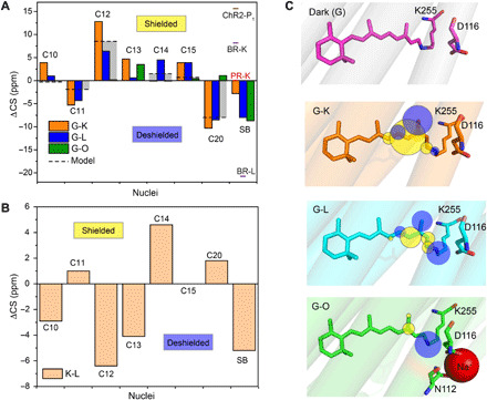
Fig. 4. Light-induced 13C retinal carbon and SB chemical shift changes.

(A) Chemical shift differences between dark state (G) and photointermediates K, L, and O are plotted for carbon atoms C10 to C15, C20, and SB. The gray bars illustrate the effect of the all-trans 13-cis isomerization as observed for the model compounds N-retinylidenebutylamine (C10 to C15) and retinal (C20) (44, 45). The 15N chemical shift of KR2 in the dark and intermediate states is compared to GPR (37), BR (28), and ChR2 (36). (B) Chemical shift difference between the K- and L-state. (C) Illustration of chemical shift changes observed at retinal carbons and SB nitrogen in the dark state [Protein Data Bank (PDB): 6REW], K-state (PDB: 6TK5), L-state (PDB: 6TK4), and O-state (PDB: 6XYT). The yellow and blue spheres indicate shielding and deshielding at the nuclei, respectively.
