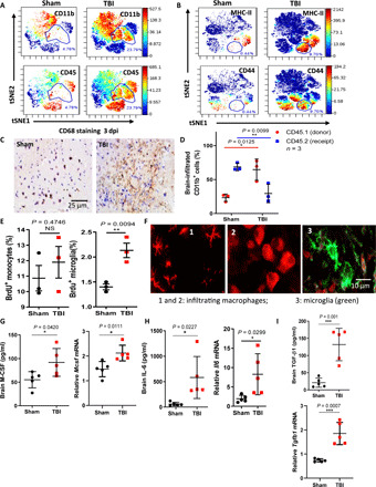
Fig. 3. Infiltration of blood-borne macrophages with microglia-like morphology and the enhanced M6T concentrations in the injured brain.

(A and B) Percentages of CD11b+, CD45+, MHC-II+, and CD44+ immune cells in sham or TBI mice brain at 3 day-post-injury (dpi) by CyTOF. (C) Representative images of immunohistochemical staining for CD68+ in sham or TBI mice at 3 dpi. (D) [CD45.1 > CD45.2] chimeric mice were reconstituted by adoptively transfer of RFP+ CD45.1 BM cells into lethally irradiated recipient CD45.2 mice. Following TBI, the percentages of CD45.2+CD11b+ or CD45.1+CD11b+ cells in the injured brain were checked by FACS. (E) 5-bromo-2′-deoxyuridine (BrdU) was injected into TBI mice at 3 dpi, and 24 hours later, cells were harvested for FACS (n = 3). (F) tdTomato+ bone marrow-derived macrophages (BMMs) were transferred into lethally irradiated recipient mice, followed by TBI. Representative images showed morphology changes of infiltrating macrophages in the injured brain sections. (G to I) Concentrations and mRNA levels of M-CSF (G), IL-6 (H), and TGF-β1 (I) in sham or TBI mice brain by ELISA or real-time polymerase chain reaction (RT-PCR) (n = 5). NS, not significant (P > 0.05); *P < 0.05, **P < 0.01, ***P < 0.001, and ****P < 0.0001. Statistical significance was determined using an unpaired Student’s t test (E and G to I) or two-way ANOVA with Sidak’s multiple comparisons test (D). tSNE, t-Distributed Stochastic Neighbor Embedding.
