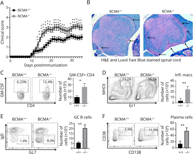
Figure 1. BCMA Deficiency Exacerbated EAE Disease Severity.
(A) EAE was induced in BCMA−/− mice and BCMA+/+ mice and scored daily for disease severity. Data are pooled from 3 experiments (n = 15–18/group). Nonparametric Mann-Whitney tests were used to determine statistical significance (*p < 0.05 and **p < 0.01). (B) Representative spinal cord sections from BCMA+/+ and BCMA−/− mice harvested at EAE day 15. Sections were stained with hematoxylin and eosin (H&E) and Luxol fast blue, and arrows indicate lesions. Dark purple color indicates infiltrating cells, and blue color indicates the myelin. The presence of (C) GM-CSF+ CD4 T cells, (D) MHCII+GR1+ inflammatory macrophages, and (E) GL7+IgD− germinal center (GC) B cells and (F) CD138+CD38+ plasma cells in the spinal cords of BCMA+/+ and BCMA−/− mice (EAE day 15) was measured by flow cytometry. Data are pooled from 2 experiments (n = 5–10/group). Error bars represent SEM, and Student t tests were used to determine statistical significance (*p < 0.05 and **p < 0.01). BCMA = B cell maturation antigen; EAE = experimental autoimmune encephalomyelitis; Ig = immunoglobulin; MHCII = major histocompatibility complex class II.

