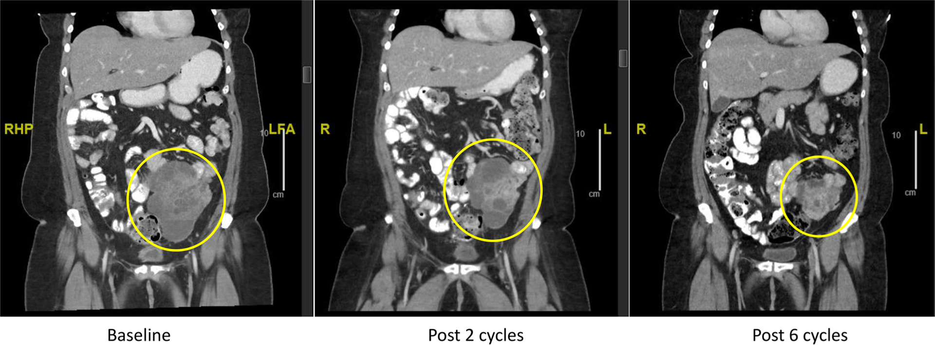
Fig. 2.

Clinical response in a patient with Sertoli–Leydig tumor harboring AKT3 amplification and STK11 R304W mutation. Computed axial tomography (CT) coronal images are shown, yellow circles highlight the index mesenteric mass; baseline (left panel), following two cycles (middle panel), following six cycles (right panel)
