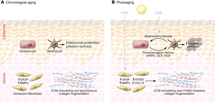
Figure 3. Differences between the chronologically aged and photoaged skin microenvironments.
A summary of key differences between chronologically aged (A) and photoaged (B) skin as described in “The photoaged microenvironment and melanoma progression.” Intrinsic aging is associated with decreases in melanocyte proliferation and melanogenesis, resulting in hypopigmentation of the epidermis. In contrast, UVR-exposed keratinocytes induce melanogenesis, thereby causing regions of hyperpigmentation clinically recognized as “sun spots.” Both the intrinsically aged and the photoaged fibroblasts of the dermal skin layer express SASP and increased production of MMPs, which leads to collagen breakdown and ECM remodeling. The mechanisms driving spontaneous collagen fragmentation in intrinsically aged skin remain largely unknown. However, in photoaged fibroblasts, this process is thought to be mediated by an increased hyaluronic acid depolymerization by HYBID and downregulation of collagen production by altered miRNAs and circRNAs.

