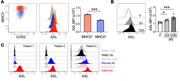
Figure 1. AXL expression is increased on human and murine macrophages after myocardial ischemia/reperfusion infarction (IRI).
(A) Cell-surface protein expression of AXL on different cardiac macrophage subsets with quantification of AXL expression on MHCIIhiCCR2– (red) and MHCIIloCCR2– (blue) macrophages. The gray histogram represents Axl–/– cardiac macrophage staining control. Data are representative of 3 independent experiments. n = 3 mice/group. ***P < 0.001 by 2-tailed, unpaired t test. (B) AXL expression on murine cardiac macrophages before (Ø) and 3 or 30 days after myocardial IRI. n = 4–7 mice/group pooled from 2 independent experiments. *P < 0.05, ***P < 0.001 by 1-way ANOVA followed by Tukey’s test. (C) Expression of AXL on peripheral blood mononuclear cells (PBMCs) or human cardiac macrophages isolated from peri-infarct or remote tissue from the explanted hearts of patients with ischemic cardiomyopathy at the time of heart transplantation. Fluorescence minus one (FMO) was used as a staining control. All data presented as mean ± SEM.

