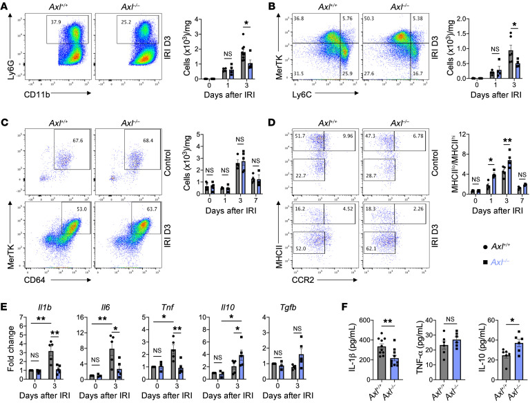
Figure 3. Macrophage AXL promotes inflammatory responses after myocardial ischemia/reperfusion infarction (IRI).
Total number of (A) neutrophils and (B) Ly6Chi monocytes within the infarcted myocardium as measured on the days indicated after IRI in Axl+/+ or Axl–/– mice. Flow plots depict events 3 days after IRI. (C) Total number of MerTK+ macrophages within the infarcted myocardium as measured on the indicated days after IRI in Axl+/+ or Axl–/– mice. (D) Ratio of MHCIIlo to MHCIIhi macrophages (MΦ) within the infarcted myocardium as measured on the days indicated after IRI in Axl+/+ or Axl–/– mice. For A–D, n = 4–5 mice/group pooled from 3 independent experiments. *P < 0.05, **P < 0.01 by 2-way ANOVA followed by Tukey’s test. (E) Gene expression of pro- and antiinflammatory mediators in whole-infarct extracts from Axl+/+ or Axl–/– mice. n = 3–5 mice/group pooled from 3 independent experiments. *P < 0.05, **P < 0.01 by 2-way ANOVA followed by Tukey’s test. (F) Serum levels of pro- and antiinflammatory mediators as measured 3 days after IRI from in Axl+/+ or Axl–/– mice. n = 6–10 mice/group pooled from 3 independent experiments. *P < 0.05, **P < 0.01 by 2-tailed, unpaired t test. All data presented as mean ± SEM.

