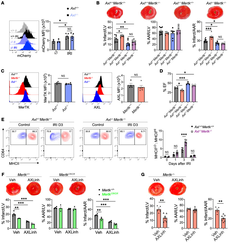
Figure 7. Divergent roles for AXL and MerTK in cardiac repair after myocardial ischemia/reperfusion infarction (IRI).
(A) Phagocytosis of apoptotic mCherry-expressing cardiomyocytes by cardiac macrophages from Axl+/+ or Axl–/– mice at baseline (Ø) or 4 hours after IRI. n = 3–5 mice/group pooled from 3 independent experiments. *P < 0.05 by 2-way ANOVA followed by Tukey’s test. (B) Percentage infarct/left ventricle (LV), percentage area at risk (AAR)/LV, and percentage infarct/AAR measured 7 days after IRI in mice lacking Mertk (Axl+/+ Mertk–/–), Axl (Axl–/– Mertk+/+), or both Mertk and Axl (Axl–/– Mertk–/–). n = 6–9 mice/group pooled from more than 3 independent experiments. *P < 0.05, **P < 0.01, ***P < 0.001 by 1-way ANOVA followed by Tukey’s test. (C) Expression of MerTK and AXL on cardiac macrophages from Axl- or Mertk-deficient mice as measured 3 days after IRI. n = 4–5 mice/group pooled from 2 independent experiments. NS, not significant by 2-tailed, unpaired t test. (D) Quantification of percentage ejection fraction (% EF) in mice 28 days after IRI. n = 4 mice/group pooled from 4 independent experiments. *P < 0.05, ***P < 0.001 by 1-way ANOVA followed by Tukey’s test. (E) Ratio of MHCIIlo to MHCIIhi cardiac macrophages within the infarcted myocardium of Axl+/+ Mertk+/+ or Axl–/– Mertk–/– mice. n = 3–5 mice/group pooled from 3 independent experiments. ***P < 0.001 by 2-way ANOVA followed by Tukey’s test. (F) Infarct measurements 7 days after IRI in Mertk+/+ or MertkCR/CR mice treated with the AXL-selective inhibitor R428 or vehicle. n = 3–4 mice/group pooled from 3 independent experiments. **P < 0.01, ***P < 0.001 by 2-way ANOVA followed by Tukey’s test. (G) Infarct measurements 7 days after IRI in Mertk–/– mice treated with the AXL-selective inhibitor R428 or vehicle. n = 5–7 mice/group pooled from 3 independent experiments. **P < 0.01 by 2-tailed, unpaired t test. All data presented as mean ± SEM.

