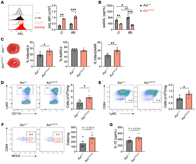
Figure 9. AXL cleavage limits adverse ventricular remodeling after myocardial ischemia/reperfusion infarction (IRI).
(A) Cell-surface expression of AXL on cardiac macrophages from wild-type (Axl+/+) or AXL cleavage–resistant (AxlCR/CR) mice before (Ø) or 3 days after IRI. (B) Serum levels of soluble AXL (solAXL) before or 3 days after IRI. For A and B, n = 3–5 mice/group pooled from 3 independent experiments. *P < 0.05, **P < 0.01, ***P < 0.001 by 2-way ANOVA followed by Tukey’s test. (C) Percentage infarct/left ventricle (LV), percentage area at risk (AAR)/LV, and percentage infarct/AAR measured 7 days after IRI in Axl+/+ or AxlCR/CR mice. n = 4–6 mice/group pooled from 3 independent experiments. *P < 0.05, **P < 0.01 by 2-tailed, unpaired t test. Total number of (D) neutrophils, (E) Ly6Chi monocytes, and (F) MHCIIhi macrophages within the infarct 3 days after IRI. Flow plots depict events 3 days after IRI. (G) Serum levels of IL-1β as measured 3 days after IRI. For D–G, n = 5 mice/group pooled from 2 independent experiments. *P < 0.05 by 2-tailed, unpaired t test. All data presented as mean ± SEM.

