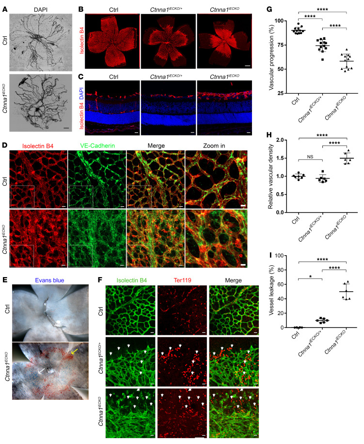
Figure 4. Conditional KO of Ctnna1 in mice causes severe retinal vascularization defects.
(A) DAPI staining of hyaloid vessels in the eyes of control and Ctnna1iECKO mice, showing that hyaloid vessel regression was markedly delayed in the eye. Scale bar: 250 μm. (B) Compared with those of littermate controls, P9 flat-mounted retinas of Ctnna1iECKO/+ mice showed delayed radial growth of the superficial vascular plexus, with moderate neovascularizations at the angiogenic front. Ctnna1iECKO retinas showed retarded vascular growth and hyperplasia of the primary vascular plexus. Vessels were stained with IB4 (red). Scale bar: 500 μm. (C) Frozen sections of retinas from P9 control, Ctnna1iECKO/+, and Ctnna1iECKO mice were costained with IB4 (red) and DAPI (blue). Scale bars: 25 μm. Vertical growth of the superficial retinal vascular plexus was delayed in Ctnna1iECKO/+ mice. In Ctnna1iECKO retinas, profound defects in vertical vascular growth into the deeper retinal layers were observed, and the vascular plexus became hyperplastic. No secondary or tertiary vessels were observed in these retinas. (D) VE-cadherin (green) and IB4 (red) staining of P9 Ctnna1iECKO and control retinas. VE-cadherin was disorganized in Ctnna1iECKO retinas. White dotted boxes indicate enlarged regions, detailed on the right. Scale bars: 25 μm and 10 μm (enlarged insets). (E) P9 flat-mounted Ctnna1iECKO retinas showed extensive leakage of Evans blue dye (white arrow) and visible, enlarged blood vessels (yellow arrow) compared with control retinas. (F) Ctnna1iECKO, Ctnna1iECKO/+, and control retinas were costained with IB4 (green) and Ter119 (red), revealing extensive leakage of erythrocytes (white arrowheads) in Ctnna1iECKO/+ and Ctnna1iECKO retinas. Scale bars: 25 μm. (G–I) Quantification of vascular progression, vascular density, and vessel leakage. Error bars indicate the SD. *P <0.05 and ****P < 0.0001, by 1-way ANOVA with Tukey’s multiple-comparison test (n ≥ 6). Experiments were performed independently at least 3 times.

