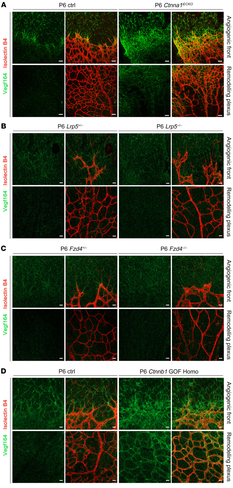
Figure 7. VEGFA distribution in Ctnna1 endothelial conditional KO, Lrp5-KO, Fzd4-KO, and Ctnnb1 GOF Homo mice.
(A) VEGF164 (green) and IB4 (red) staining of P6 control and Ctnna1iECKO retinas. Abnormal distribution and elevated expression of VEGF164 expressed by both astrocytes and ECs were observed in the angiogenic front and remodeling plexus of Ctnna1iECKO retinal vessels. (B and C) In P6 Lrp5+/–, Lrp5–/–, Fzd4+/–, and Fzd4–/– retinas, VEGF164 was localized normally in the angiogenic front and absent in the remodeling plexus. Scale bars: 25 μm. (D) In P6 control and Ctnnb1 GOF Homo retinas, abnormal distribution and elevated VEGF164 expression by both astrocytes and ECs were observed in the angiogenic front, whereas in the remodeling plexus, only endothelium-derived VEGF164 expression was elevated. Experiments were performed independently at least 3 times.

